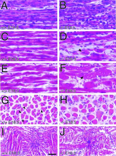
Figure 2.
Deformed trio−/− embryonic skeletal myofibers. Shown are H&E-stained sagittal sections of matched thoracic paraspinal skeletal muscle from trio+/+ (A, C, and E) and trio−/− (B, D, and F) embryos at E14.5 (A and B), E16.5 (C and D), and E18.5 (E and F) of development. From E14.5 to E18.5 there is an increase in the number of abnormal large rounded myofibers (see arrows). (G and H) H&E staining of E18.5 trio+/+ (G) and trio−/− (H) coronal sections of the same muscles shown in A–F, demonstrating the abnormally large and morphologically unusual trio−/− myofibers. Arrows in G indicate the location of some secondary myofibers in trio+/+ embryos. (I and J) H&E staining of E18.5 trio+/+ (I) and trio−/− (J) coronal sections of tongue muscle. (Scale bar represents 25 μm for panels A–H, and 50 μm for I and J.)

