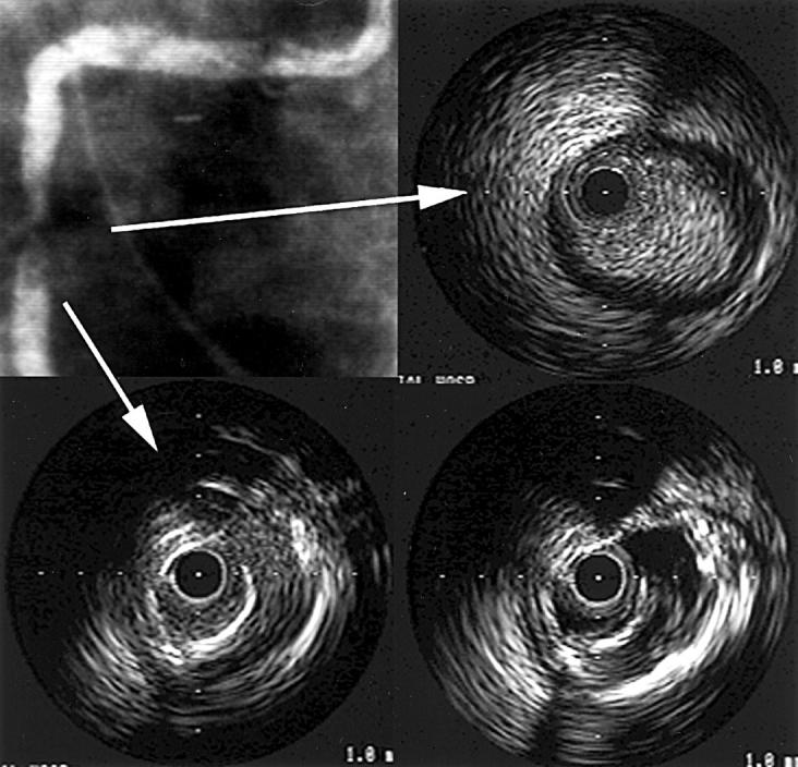
Figure 1 .

Coronary angiography and intravascular ultrasound (IVUS) images—1. The coronary angiography and IVUS images of a patient with acute myocardial infarction in the right coronary artery territory. (A, top left) Coronary angiography shows the right coronary artery with 99% stenosis in the mid-portion. (B, top right) A plain IVUS image of proximal stenosis. This shows bright speckled echo material. (C, bottom left) A plain IVUS image with suspected plaque dissection. (D, bottom right) Negative contrast image of (C). This reveals the plaque dissection at 3 o'clock more clearly than the plain image.
