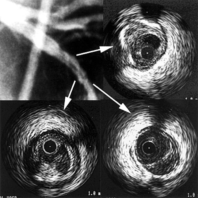
Figure 2 .

Coronary angiography and intravascular ultrasound (IVUS) images—2. The coronary angiography and IVUS images of a patient with acute myocardial infarction in left anterior descending coronary artery (LAD) territory. (A, top left) Coronary angiography shows filling defect in the mid-portion of the LAD. (B, top right) IVUS image of low echoic thrombus. In concordance with the angiographic findings, a low echoic mass representing thrombus can be seen at 12 to 2 o'clock. (C, bottom left) IVUS image with subtle dissection at 6 o'clock. (D, bottom right) IVUS image representing echolucent area at 4 to 6 o'clock; a low echoic lesion can be seen in the eccentric plaque.
