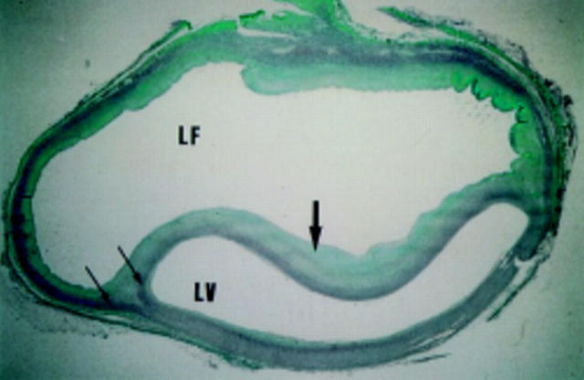
Figure 2 .
Histological section (Mason's technique) from a patient with aortic dissection. Muscle is stained in red and collagen in green. The aortic media (stained in red) is partitioned in two (arrows); one forms part of the dissection flap, the other forms the outer wall of the false channel. Large arrow indicates the dissection flap. LF, false lumen; LV, true lumen.

