Figure 2: .
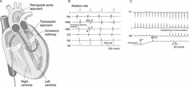
(A) Schematic drawing of the two approaches which are available to ablate left sided accessory pathways. The retrograde aortic approach involves inserting the ablation catheter into the femoral artery and crossing the aortic valve to enter the left ventricle. The ablation catheter is positioned against the ventricular aspect of the mitral annulus. The transseptal approach involves crossing the interatrial septum and positioning a long transeptal sheath into the left atrium. The ablation catheter is then passed through the sheath and positioned against the atrial aspect of the mitral annulus at the site of the location of the accessory pathway. (B) The electrogram characteristics of a typical successful ablation site of an accessory pathway (ABL) are shown. Also shown are the surface leads I and V6 and intracardiac recordings obtained from the high right atrium (RA), the right ventricle apex (RV), the electrode catheter positioned to record a His bundle (HBE), and an electrode catheter positioned in the coronary sinus os (CS). The surface leads show a short PR interval and slurring of the upstroke of the QRS complex, which are characteristic of the pre-excitation pattern observed in patients with the Wolff-Parkinson-White syndrome. The interval from the His bundle recording (H) to onset of the QRS complex is less than 50 ms, confirming the presence of pre-excitation. At the successful ablation site, the ventricular electrogram (V) occurs very early relative to the onset of the QRS complex. Also observed is a discrete deflection between the atrial (A) and the ventricular components of the electrogram recorded at the ablation site, which is suggestive of an accessory pathway potential. (C) The disappearance of pre-excitation several seconds after onset of radiofrequency energy delivery during catheter ablation of an accessory pathway. Shown are the surface leads V1, I, V6, and the temperature recorded from the ablation catheter. The temperature recorded from the ablation electrode increases from 37°C to 66°C within two seconds of radiofrequency energy delivery. Pre-excitation resolves several seconds thereafter.

