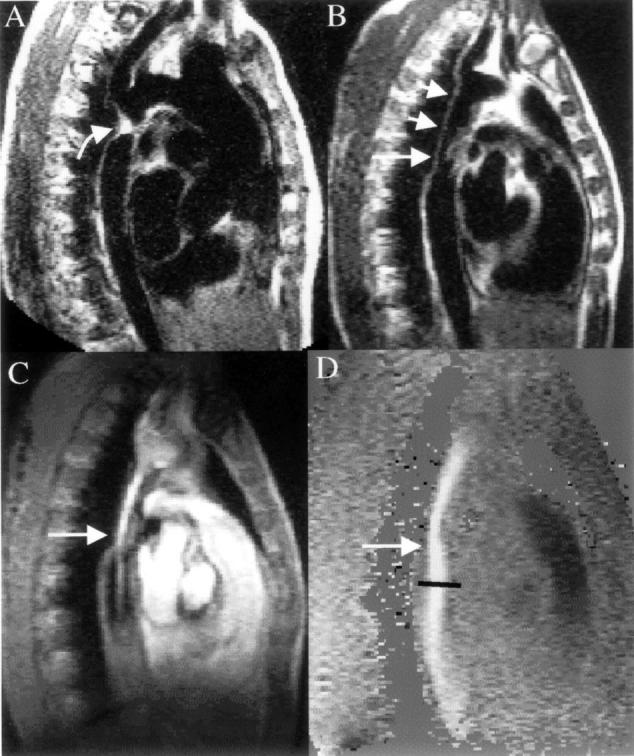
Figure 1: .

CMR in a patient with coarctation. (A) The preoperative spin echo image in an oblique sagittal plane shows the entire thoracic length of the aorta, and the coarctation (curved arrow) just distal to an enlarged left subclavian artery. (B) Appearance of the same region with spin echo imaging some years after repair of the coarctation site with a Dacron graft (short arrows). There is narrowing of the distal end of the graft and this is clearly seen in C with the systolic frame of the gradient echo cine showing bright signal within the graft from increased velocities and flow enhancement. Immediately distal to the graft narrowing (straight arrow) a bright jet is seen exiting into the normal descending aorta surrounded by dark areas which are caused by signal loss from turbulence. The velocity map D, from exactly the same plane as C, shows intense white colouration and a peak velocity measured at 3 m/s (36 mm Hg pressure gradient).
