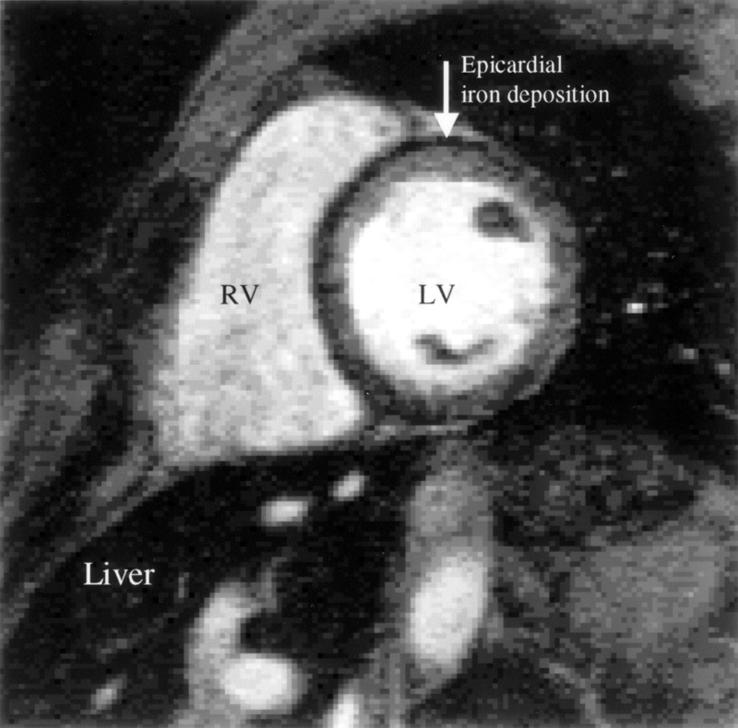
Figure 7: .
Example of iron deposition in a patient with thalassaemia. The dark epicardial rim of iron is arrowed. Note that liver deposition is very heavy, and the liver is therefore black. The signal loss occurs because of disturbances in the relaxation parameters of the tissues brought about by the iron causing alterations in the local magnetic field. There is very poor correlation between iron deposition in the liver and the heart, which prevents adequate management of the cardiac complications of myocardial iron overload (arrhythmia, heart failure, and death) from liver biopsy results. RV, right ventricle; LV, left ventricle. Reproduced from Rajappan et al, Eur J Heart Failure 2000;2:241-52, with permission of the publisher.

