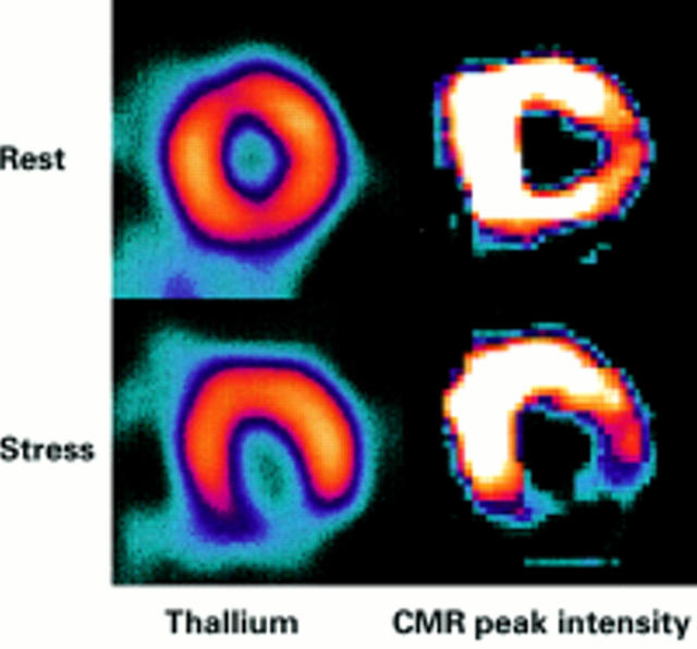
Figure 9: .
Comparison of thallium (left column) and CMR (right column) perfusion imaging in a patient with right coronary stenosis and inferior reversible ischaemia. The CMR images are parametric maps which are colour coded to appear similar to the thallium scan. Each pixel in the image represents the relative time to peak enhancement and bright colour indicates faster contrast wash-in and therefore better perfusion. The defect in the CMR scan is very similar in size and intensity to the thallium scan.

