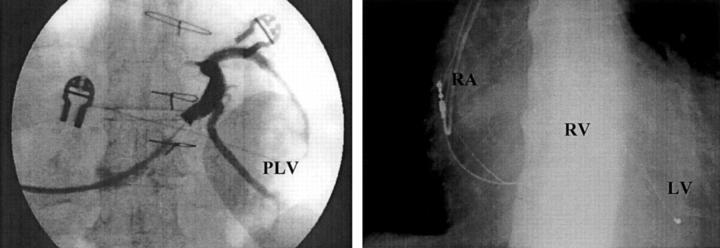
Figure 4 .
In this coronary sinus angiogram (left) there was a large posterolateral vein. However, the angle it makes with the coronary sinus may make it difficult to reach. In this case a very thin lead, with side wire technology, could be introduced into the vein (chest x ray on the right). LV, left ventricular lead; PLV, posterolateral vein; RA, right atrial lead; RV, right ventricular lead.

