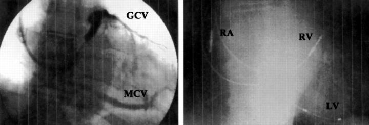
Figure 5 .
It can be seen on this coronary sinus angiogram (left) that there was no lateral or posterolateral vein, so that a choice had to be made between the great cardiac vein and the mid-cardiac vein. In this case the left ventricular lead was placed in the mid-cardiac vein, as can be seen on the chest x ray (right). GCV, great cardiac vein; LV, left ventricular lead; MCV, mid-cardiac vein; RA, right atrial lead; RV, right ventricular lead.

