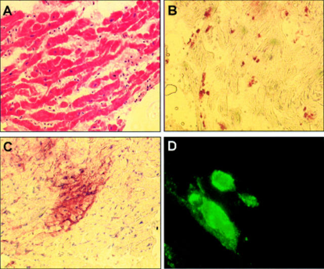
Figure 2 .
Endomyocardial biopsy results. (A) Haematoxylin and eosin staining, ×200. (B) CD45 immunostaining with horseradish peroxidase signal detection, ×200. Note the abundance and foci of inflammatory cells. (C) Immunostaining for the enteroviral VP-1 antigen with horseradish peroxidase signal detection, ×200. Note the strong sarcolemmal staining pattern in a group of cardiomyocytes. (D) VP-1 immunofluorescence staining of a mouse heart experimentally infected with coxsackievirus B3, ×400. The virally infected cells show a strong signal similar to the pattern described in C.

