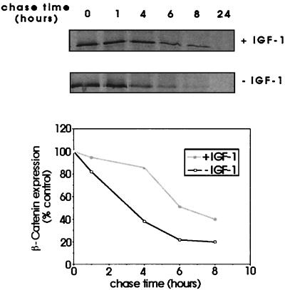
Figure 5.
Analysis of effects of IGF-1 signaling on stability of β-catenin protein. Subconfluent C10 cells were pulsed with 35S-Promix and chased with medium containing an excess of cold methionine/cysteine for the indicated times. Cells were lysed and immunoprecipitated for β-catenin. The results were analyzed by densitometry and expressed graphically as a percentage of the value at time 0 h. The figure shows results of a single experiment, which was repeated once with similar results.

