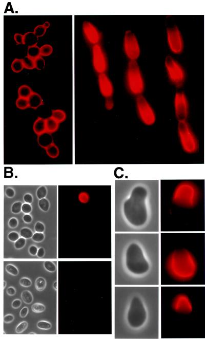
Figure 5.
Adhesion is associated with the presence of Flo11p and Fig2p on the cell surface. The Flo11p and Fig2p proteins were tagged with HA (see Materials and Methods). Yeast cells were harvested, fixed, treated with mouse anti-HA antibody, and stained with Cy3-conjugated goat anti-mouse IgG antibody. (A) Haploid yeast cells (strain WY423) grown on synthetic complete medium show uniform staining of Flo11–3HA (Left). Diploid pseudohyphal cells (strain WY453) grown on SLAD (with 0.2 mM l-histidine hydrochloride added) often show polarized staining (Right); however, some show uniform staining (see filament on left). (B) Most diploid yeast cells (WY453) fail to stain Flo11p whether they are grown on synthetic complete medium (Upper) or SLAD (Lower). (C) Haploid yeast cells treated with α factor form mating projections (Left). Only the projections show Fig2–3HA staining (strain WY427) (Top Right). Flo11–3HA appears on the body of the cell and not in the projection (strain WY423) (Middle Right). Flo11–3HA induced with Gal appears only in the projection (strain WY491) (Bottom Right).

