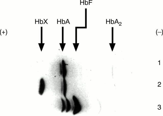
Figure 3 Isoelectric focusing gel electrophoresis of a control subject, cord blood, and the patient with Hb Takamatsu. Lane 1, electrophoretic pattern of control subject; lane 2, electrophoretic pattern of Hb Takamatsu; lane 3, electrophoretic pattern of cord blood. Abnormal haemoglobin migrating faster than HbA1 is indicated by HbX.

An official website of the United States government
Here's how you know
Official websites use .gov
A
.gov website belongs to an official
government organization in the United States.
Secure .gov websites use HTTPS
A lock (
) or https:// means you've safely
connected to the .gov website. Share sensitive
information only on official, secure websites.
