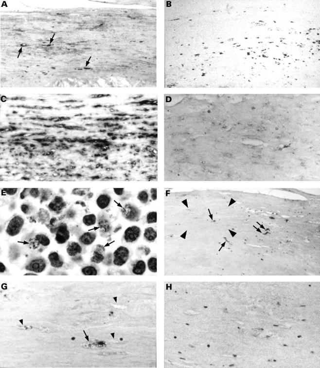
Figure 1 Representative photomicrographs of an area with atherosclerotic changes in consecutive sections of the carotid artery specimen of case 11 (A–D, and F–H), and of HEp2 cells infected with Chlamydia pneumoniae (E). Several cells in this atherosclerotic area were immunoreactive for C pneumoniae membrane protein (A; some positive cells indicated with an arrow). These cells are part of the population of cells that immunostained for macrophage marker CD68 (B). In situ hybridisation reactivity for eukaryotic 28S rRNA (C) demonstrated the suitability of the specimen for hybridisation experiments. However, C pneumoniae DNA was not detected by in situ hybridisation (D). Fragmented DNA of C pneumoniae in a section of HEp2 cells infected with C pneumoniae pretreated with DNAse I and then processed in the in situ DNA nick end labelling (TUNEL) assay was visible as uniformly sized dots in clearly recognisable inclusions (E; arrows) next to TUNEL stained nuclei of the HEp2 cells. Uniformly sized dots stained using the TUNEL assay were also found in the carotid artery specimen (F and G; arrows) in a cell type in which C pneumoniae membrane protein was also observed. An enlargement of the area enclosed by the arrowheads in (F) is shown in (G). Note that cells similar in morphology to TUNEL reactive cells were also not reactive in the TUNEL assay (G; arrowheads). These vacuolated cells are part of the population of cells that immunostained for macrophage marker CD68 (B). No staining was observed without transferase in the TUNEL reaction mixture (H). Counterstaining, panels A–D, nuclear fast red; panels E–H, Mayer's haematoxylin. Original magnifications, x31 (A, B, and F), x62 (C), x125 (D, G, and H), and x250 (E).
