
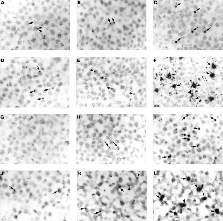
Figure 1 The detection of Chlamydia pneumoniae in paraffin wax embedded sections of formalin fixed HEp2 cells infected with a multiplicity of infection of 0.1 inclusion forming units of C pneumoniae. Cells were fixed at 21 (A, D, G, and J), 29 (B, E, H, and K), and 46 (C, F, I, and L) hours after infection. Sections were immunostained for C pneumoniae membrane protein (A–C), LPS (D–F; with monoclonal antibody 16.3B6), or hsp60 (G–I), or hybridised for 16S rRNA (J–L). In the early stages of the developmental cycle of C pneumoniae, up to 29 hours post infection, only very small inclusions were seen (A, B, D, E, H, J, and K; arrows). No immunostaining was observed for hsp60 at 21 hours after infection (G). Inclusions were up to 10 times larger at a later stage of the developmental cycle (C, F, I, and L; arrows). Membranes of non-infected cells (F; large arrowheads) and vesicle-like structures (F; small arrowheads) were also immunostained for LPS. Counterstaining, nuclear fast red. Original magnification, x125 (A–L).