
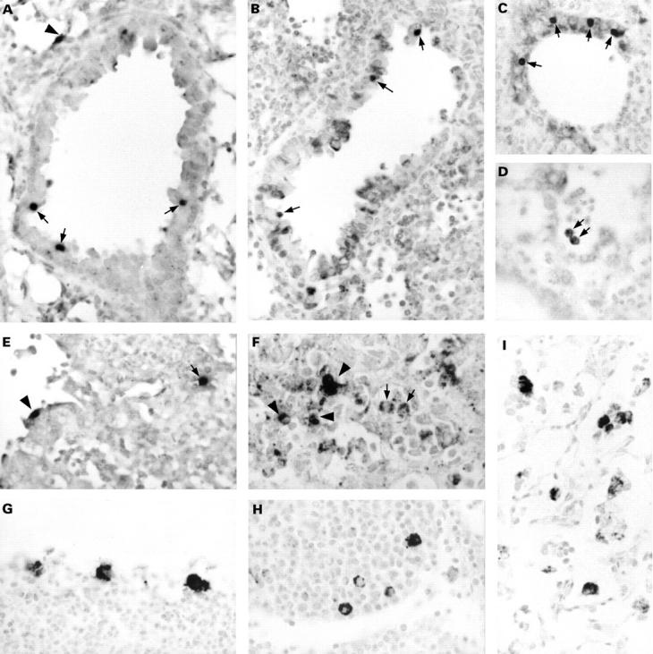
Figure 2 The detection of Chlamydia pneumoniae in paraffin wax embedded sections of formalin fixed lungs of mice intranasally infected with C pneumoniae. Results are from mice sacrificed at two (A–F) and 21 (G–I) days after infection. Sections were immunostained for membrane protein (G), lipopolysaccharide (B, F, and I; with monoclonal antibody 16.3B6), or hsp60 (C, D, and H), or hybridised for DNA (A) or 16S rRNA (E). In the early stages of infection, clearly recognisable inclusions were observed in bronchus epithelial cells (A–C; arrows) and in alveolar epithelial cells (A and E; arrowhead). Alveolar macrophages showed a vacuolised immunostaining pattern (D and F; arrows). Infiltrate cells were positive by immunostaining (F; arrowheads) and by in situ hybridisation (E; arrow). In the later stages of infection, macrophages in bronchus associated lymphoid tissue (G and H) and alveolar macrophages (I) showed a granular immunostaining pattern dispersed throughout their cytoplasm. Counterstaining, nuclear fast red. Original magnification, x125 (A–I).