

Abstract
A 7 cm diameter presacral tumour, not related to the intrapelvic organs, was found in a 51 year old woman. The needle biopsy showed a poorly differentiated large cell carcinoma. The patient died of urosepsis after chemotherapy. Postmortem examination revealed no other primary or metastatic tumour. Histological examination of the presacral tumour showed a large cell carcinoma with a trabecular pattern and strong immunoreactivity for neuroendocrine markers. The tumour was finally classified as a primary large cell neuroendocrine carcinoma of the presacral region.
Key Words: neuroendocrine carcinoma • presacral region
Full Text
The Full Text of this article is available as a PDF (167.5 KB).
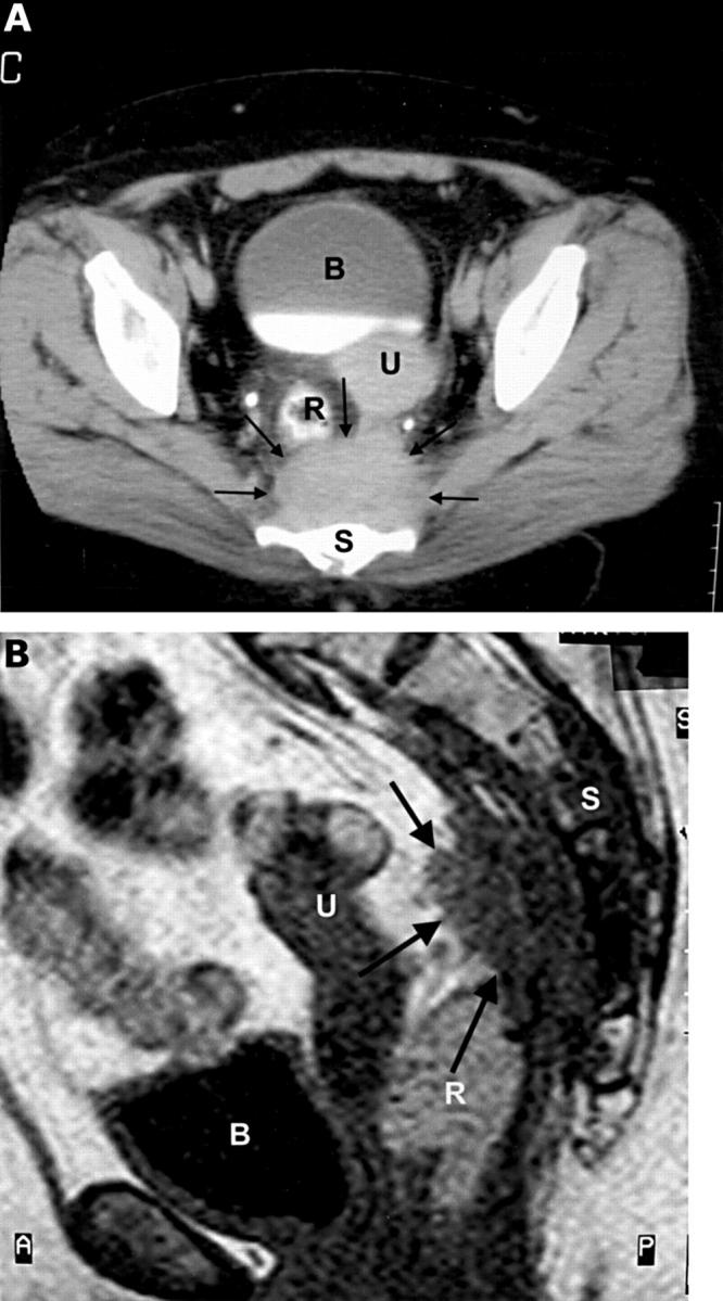
Figure 1 (A) Axial computed tomography scan cut through the pelvis depicting the presacral tumour (arrows). Notice the adjacent structures: B, bladder; R, rectosigmoid; U, uterus; S, sacrum. (B) Sagittal T1 weighted magnetic resonance image through the pelvis. Notice the invasive growth of the tumour (arrows) in the sacrum (S), as demonstrated by the low signal intensity of the involved vertebral bodies. Notice adjacent structures: B, bladder; U, uterus; R, rectum.
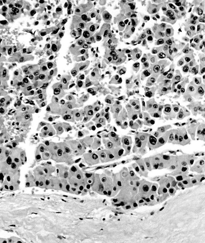
Figure 2 Microscopic picture of the tumour (postmortem specimen) showing a trabecular pattern with large sized cells that possess vesicular nuclei with prominent nucleoli (haematoxylin and eosin; original magnification, x250).

Figure 3 Immunohistochemical stain of the tumour. (A) Staining for chromogranin A: notice the perineural tumour growth. (B) Staining for cytokeratin, highlighting the trabecular growth pattern (original magnification, x100).