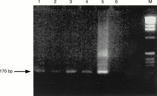
Figure 1 PCR amplification of four samples. The samples were treated with proteinase K, amplified by PCR using human cytomegalovirus (CMV) specific primers, electrophoresed in 2% agarose gel, stained with ethidium bromide, and visualised under UV light (320 nm). Lanes 1–4, positive samples; lane 5, positive control (strain AD169 of human CMV); lane 6, negative control; and lane M, molecular weight marker. The arrow shows a 170 bp fragment.

An official website of the United States government
Here's how you know
Official websites use .gov
A
.gov website belongs to an official
government organization in the United States.
Secure .gov websites use HTTPS
A lock (
) or https:// means you've safely
connected to the .gov website. Share sensitive
information only on official, secure websites.
