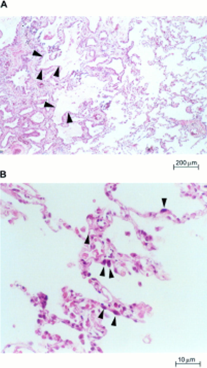
Figure 1 Lung parenchyma of patients with usual interstitial pneumonia. (A) Characteristic variation in histological appearance from one area to another. Dense collagen deposition in the lung parenchyma is seen on the left, and patchy areas containing normal or nearly normal alveoli are present nearby (right and bottom centre). There is a zone of microscopic honeycomb change at the middle left (arrowheads) that is characterised by mucin filled, enlarged air spaces separated by fibrosis (haematoxylin and eosin staining; magnification, x100). (B) High magnification view of the surviving zones of alveoli showing marginated masses of chromatin within shaped, rounded alveolar cells with eosinophilic cytoplasm (arrowheads), suggestive of type II pneumocytes (haematoxylin and eosin staining; magnification, x400).
