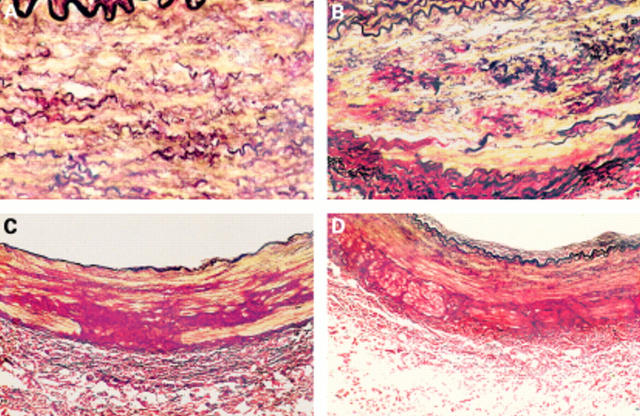
Figure 8 (A) Extracranial vertebral artery transverse section from a 17 year old subject, showing mild to moderate elastic fibre fragmentation (E1–E2). (B) Extracranial vertebral artery section from a 33 year old subject. Note the advanced elastic fibre fragmentation (E3) and severe collagen scarring (C3). (C) Transverse vertebral artery section from the upper cervical loop segment of a 30 year old subject. Note the very dense distribution of collagen extending from the adventitia into the media. (D) Transverse vertebral artery section from the upper cervical loop segment of a 33 year old subject. Again, note the unusual arrangement of peripheral collagen extending from the adventitia (all stained with picro-sirius red).

An official website of the United States government
Here's how you know
Official websites use .gov
A
.gov website belongs to an official
government organization in the United States.
Secure .gov websites use HTTPS
A lock (
) or https:// means you've safely
connected to the .gov website. Share sensitive
information only on official, secure websites.
