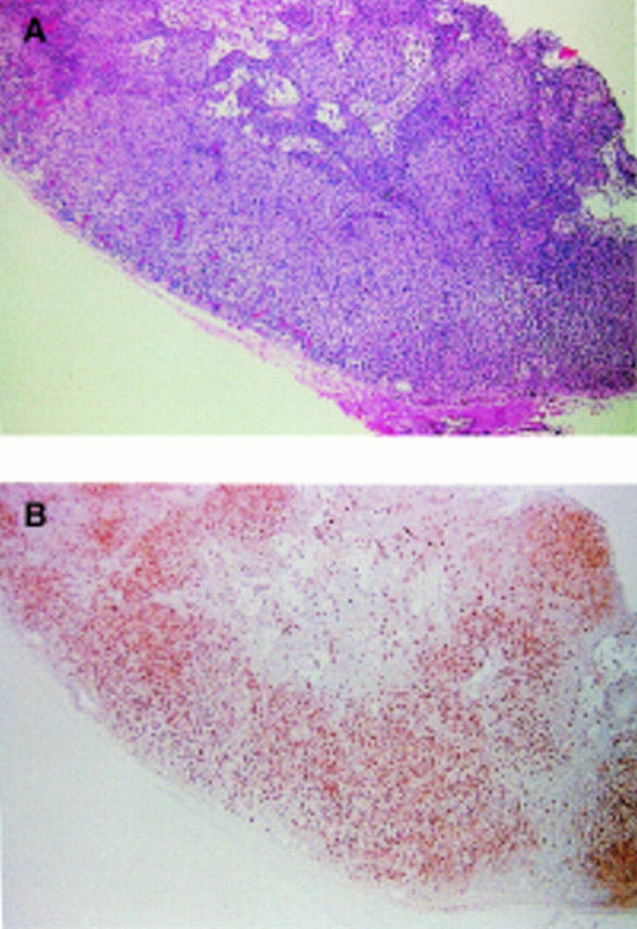
Figure 4 Lymph node biopsy in Omenn's syndrome (courtesy of B Angus). (A) Haematoxylin and eosin stained preparation. Note the absence of germinal centres and the replacement of the paracortex by a diffuse infiltrate of large cells with abundant pale cytoplasm. (B) Immunostaining for S100 protein. Note the positive labelling of almost all the cells in the cortex. This indicates infiltration of the node by a population of interdigitating reticulum cells .
