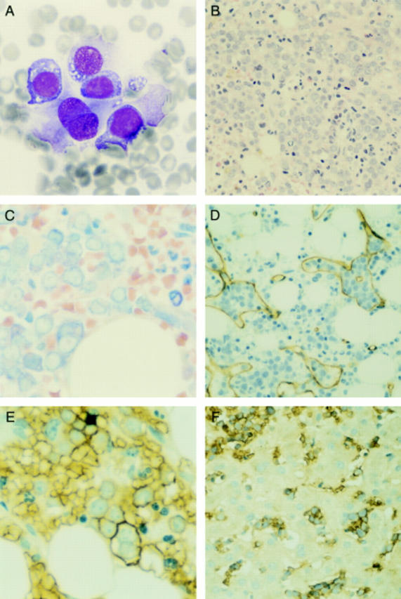
Figure 1 Case 1. (A) Bone marrow aspirate smear showing large primitive cells with vacuolated cytoplasm. (B) The primitive cells comprise about 80% of the cells of the bone marrow biopsy section, but interspersed maturing myeloid forms are also present (haematoxylin and eosin stained). (C) The primitive cells have basophilic, vacuolated cytoplasm on Giemsa stain; note the maturing myeloid forms in the upper right section. (D) The primitive forms cluster within bone marrow sinuses, as shown by the CD34 immunohistochemical stain highlighting endothelial cells. (E) The immature cells are immunoreactive for ß-sialoglycoprotein; note one binucleate erythroblast. (F) A liver biopsy shows similar cells, immunoreactive for ß-sialoglycoprotein, within hepatic sinusoids.
