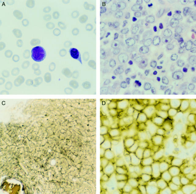
Figure 2 Case 2. (A) Peripheral blood smear showing an immature cell with basophilic vacuolated cytoplasm and a nucleated erythroid cell. (B) Bone marrow biopsy section showing sheets of large cells with vesicular nuclei and prominent nucleoli (haematoxylin and eosin stained). (C) The marrow shows greatly increased reticulin deposition (Gordon and Sweet's silver stain). (D) The immature cells are uniformly immunoreactive for glycophorin C.

An official website of the United States government
Here's how you know
Official websites use .gov
A
.gov website belongs to an official
government organization in the United States.
Secure .gov websites use HTTPS
A lock (
) or https:// means you've safely
connected to the .gov website. Share sensitive
information only on official, secure websites.
