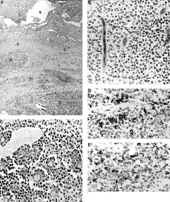
Figure 2 (A) Low power photomicrograph of the tumour (S) showing the fibrous pseudocapsule (C) and the ectopic pancreatic tissue (P). Tumour cells are arranged in (B) solid sheets or (C) form pseudopapillary structures around the blood vessels. Two characteristic immunophenotypic features were: (D) the strong, focal KL-1 positivity, and (E) the focal, granular or dot-like α-1-antichymotrypsin positivity.
