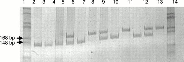
Figure 1 Sample 12% (wt/vol) acrylamide gel depicting the three p53 codon 72 genotypes detected in the patients with skin cancer and the control population. Lanes 1 and 14 contain molecular weight markers (HaeII digested pBluescript). Lanes 2, 3, 6, 9, and 11 show the single 144 bp band amplified from individuals who are arginine homozygotes for the codon 72 polymorphism. Lanes 7, 10, and 13 show the single 171 bp band amplified from the p53 gene of individuals who are proline homozygotes for the codon 72 polymorphism. Lanes 5, 8, and 12 show both the 144 bp arginine band and the 171 bp proline band amplified from the p53 gene of heterozygous individuals. The gel was stained with ethidium bromide and visualised under UV light.

An official website of the United States government
Here's how you know
Official websites use .gov
A
.gov website belongs to an official
government organization in the United States.
Secure .gov websites use HTTPS
A lock (
) or https:// means you've safely
connected to the .gov website. Share sensitive
information only on official, secure websites.
