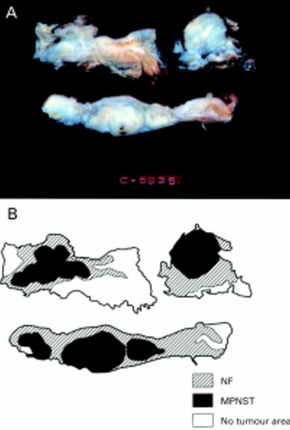
Figure 1 (A) Cut surface of the resected tumour in case 3. Section shows a multinodular solid and white mass. The central firm portion blends with the surrounding subcutaneous fat and muscular tissue. (B) Topographical distribution of histological features of both malignant peripheral nerve sheath tumour and neurofibroma in case 3.
