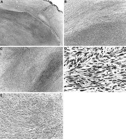
Figure 3 (A–D) Histological picture exhibiting the features of neurofibroma and malignant peripheral nerve sheath tumour (MPNST) (case 4). (A) The MPNST area is surrounded by a neurofibromatous area. Densely cellular fascicles alternate with hypocellular zones in the MPNST area (haematoxylin and eosin stained; original magnification, x12). (B) The tumour is composed of interlacing bundles of elongated cells with wavy nuclei, without atypism in the neurofibroma (top), and with atypism in the MPNST (bottom) (haematoxylin and eosin stained; original magnification, x20). (C) The tumour is composed of wavy spindle cells arranged in fascicles and with wavy nuclei. Densely cellular fascicles alternate with hypocellular zones (haematoxylin and eosin stained; original magnification, x25). (D) The nuclei of some tumour cells are wavy or buckled. Mitotic figures are seen frequently (haematoxylin and eosin stained; original magnification, x570). (E) Histological picture exhibiting the features of neurofibroma (case 4). The tumour is made up of interlacing bundles of elongated cells with wavy nuclei, without atypism, associated with wire-like strands of collagen (haematoxylin and eosin stained; original magnification, x125).
