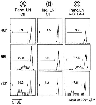
Figure 1.
No detectable effect of anti-CTLA-4 treatment on division of transferred BDC2.5 T cells in the PLNs. CFSE-labeled splenocytes from juvenile BDC2.5/NOD mice were transferred into adult Cαo/o/NOD animals that had been treated with anti-CTLA-4 mAb or PBS 2 h before transfer. PLNs and inguinal lymph node were removed from the recipients at the indicated times after transfer, and cells were stained with mAbs against CD4 and Vβ4 (the trangene-encoded TCR β-chain). Shown are histograms of CFSE staining for gated CD4+Vβ4+ cells. The reduction in CFSE staining intensity signifies that cell proliferation has taken place, and the proportion of divided cells is indicated.

