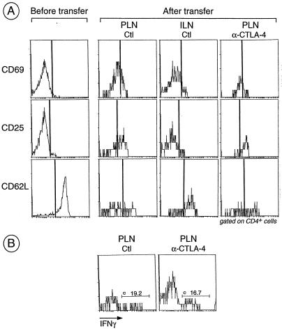
Figure 2.
Activation of BDC2.5 T cells after transfer into anti-CTLA-4-treated and control recipients. (A) Splenocytes from juvenile BDC2.5/NOD mice were pooled and an aliquot was stained for CD4, CD8, and early (CD25 and CD69) and late (CD62L) activation markers. Cells were transferred into adult Cαo/o/NOD mice that were treated with anti-CTLA-4 as described in the legend to Fig. 1. At day 3, single-cell suspensions from PLNs and inguinal lymph nodes were stained for CD4, CD8, and various activation markers; histograms gated on CD4+ cells are shown. (B) Same as in A except intracellular staining for IFN-γ was performed in place of staining for activation markers.

