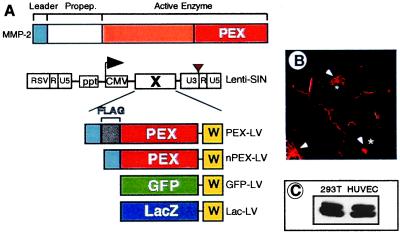
Figure 1.
Lentiviral vectors used for PEX delivery and transduction of endothelial cells with lentiviral vectors. (A) Schematic structure of MMP-2 (Upper) and lentiviral vectors (Lower). The lentiviral vectors contain the following features. The U3 element of the 5′ LTR is replaced by a Rous sarcoma virus promoter (RSV) that drives expression of the vector transcripts in the packaging cells. The 3′ LTR contains a SIN mutation (brown triangle) to ensure self-inactivation in the target cell. Expression of the transgene (X) is driven by the internal cytomegalovirus (CMV) promoter. The difference between nPEX-LV and PEX-LV is the inclusion of a FLAG-tag (gray) in the latter. W, posttranscriptional regulatory element of woodchuck hepatitis virus (yellow); ppt, polypurine tract. (B) Analysis of PEX expression by HUVECs transduced with PEX-LV using Abs directed against the FLAG-tag. Arrowheads, Golgi apparatus; *, nucleus. (C) Western blot analysis of PEX expression in 293T (left lane) cells stably expressing PEX and HUVECs (right lane) transduced with PEX-LV.

