
Fig. 1. In vitro cleavage of SK1 by cathepsin B.
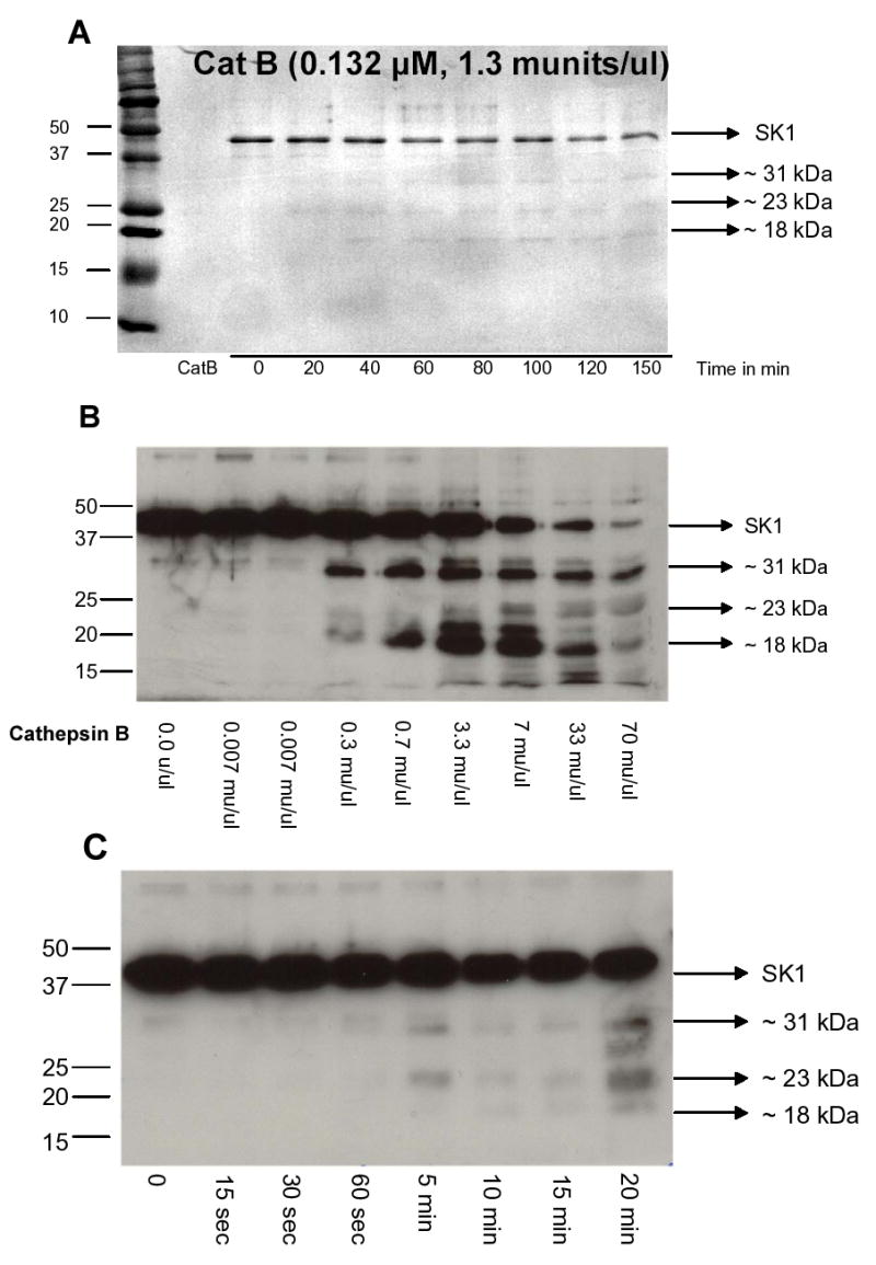
(A) Recombinant SK1 was incubated with recombinant cathepsin B for various times in a cleavage assay as described in “Materials and Methods”. The products of the reaction were run on an SDS-PAGE, transferred to PVDF membrane and stained with Coomassie Brilliant Blue. Several cleavage fragments were identified. (B) Dose response of recombinant cathepsin B incubated with 500 ng of recombinant SK1. The products of the cleavage reaction were analyzed by Western blotting using an SK1 antibody. (C) Time course of recombinant cathepsin B (1.3 munits/ul) incubated with 500 ng of recombinant SK1. The products of the cleavage reaction were analyzed by Western blotting using an SK1 antibody.