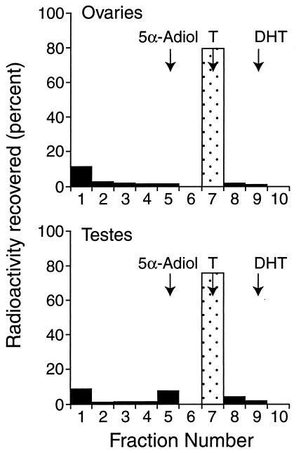
Figure 1.
TLC of the metabolites after the incubation of tissue fragments of testes or ovaries with tritiated testosterone. The positions of the standards on the plate are indicated by arrows. The data from one of three experiments are shown. Fraction 1 represents the origin—the metabolite at the origin in both the ovarian and testicular samples was not identified. T, testosterone.

