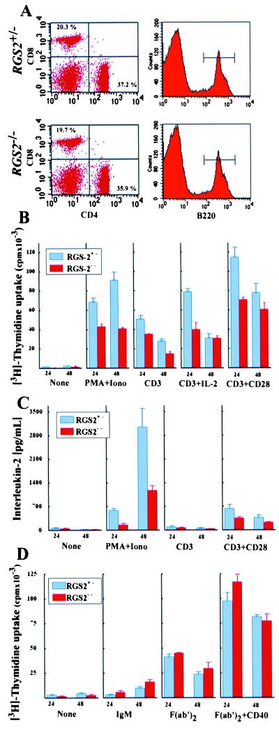
Figure 2.
Impaired proliferation and IL-2 production by rgs2−/− T cells. (A) Normal populations of CD4+ and CD8+ T cells and B220+ B cells in lymph nodes of rgs2−/− mice. Numbers in each quadrant represent percentages of each subset. (B) Proliferation. Lymph node T cells (2 × 10⁵/well) from rgs2−/− and rgs2+/− mice were activated by using PMA [10 ng/ml]+Ca+2-ionophore [100 ng/ml], TCR stimulation [anti-CD3α mAb (0.1 μg/ml)] with and without CD28 costimulation [anti-CD28 mAb (0.02 μg/ml)] or IL-2 [50 units/ml]. Results are shown as mean [3H]thymidine uptake ± SD. Difference in proliferation between rgs2+/− and rgs2−/− T cells was statistically significant (t test, P < 0.05). (C) IL-2 production. Lymph node T cells were activated as above, and IL-2 was measured by ELISA at 24 and 48 h poststimulation. Mean values of IL-2 production ± SD are shown. (D) Activation of B cells. Purified splenic B cells (1 × 10⁵/well) were incubated in triplicate for 24 and 48 h in medium alone (None) or medium containing anti-IgM (20 μg/ml), anti-IgM (Fab′)2 (15 μg/ml) with or without anti-CD40 (5 μg/ml). Results are shown as mean [3H]thymidine uptake ± SD.

