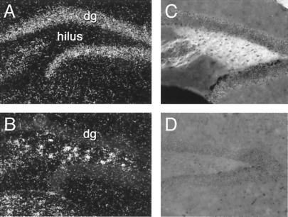
Figure 3.
Expression of c-Ret and GFRα2 mRNAs and of GFRα2 immunoreactivity in the dentate gyrus. Dark-field photomicrographs showing expression of GFRα2 (A) and c-Ret (B) mRNAs in naive C57BL/6 mice. Note the high expression of GFRα2 mRNA in granule cells (A) and of c-Ret in hilar interneurons (B). Also note that hilar interneurons do not express GFRα2 mRNA (A). C and D show the high level (C) and absence (D) of GFRα2 immunoreactivity in the hilus of wild-type (C) and knock-out (D) mice.

