Figure 4.
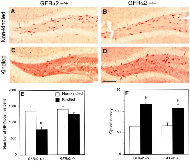
Down-regulation of NPY immunoreactivity in hilar interneurons after test stimulations in kindled animals is not observed in GFRα2 −/− mice. Color photomicrographs showing NPY immunoreactivity in the dentate hilus of non-kindled (A and B) and kindled animals (C and D). (E) The number of NPY-positive neurons; (F) the optical density of NPY immunoreactivity in the hilus of nonstimulated or kindled GFRα2 +/+ and −/− mice. Note the decrease of NPY-positive neurons in the hilus of kindled, wild-type animals (C), with no alterations in the knock-out mice (D), as compared with corresponding controls (A and B, respectively). Mean ± SEM. *, P < 0.05, Student's unpaired t test (n = 10 for GFRα2 +/+ and n = 7 for −/− mice). Bar = 160 μm.

