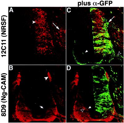
Figure 2.
Morphology of NRSF-misexpressing neurons identified by coexpression of tauGFP. Embryos were electroporated with the NTG expression construct that encodes an NRSF-IRES-tauGFP cassette. A and C show a single section double-stained with antibodies to NRSF (A) and GFP (C) visualized to display either NRSF expression alone (A) or both NRSF and tauGFP expression (C). B and D show an adjacent section double-stained with antibodies to Ng-CAM (B) and GFP (D), visualized in a similar manner. Many cells expressing exogenous NRSF show GFP staining (C); cells expressing GFP but not NRSF are rare. NRSF-overexpressing sensory neurons in the DRG (A, arrow) send processes into the dorsal spinal cord (C, arrow; see also Fig. 3E, arrow), whereas motor neurons extend processes out of the ventral root (C, open arrowhead). Expression of NRSF from this nonintegrating expression plasmid also represses Ng-CAM expression in commissural (B, arrow) and sensory (B, arrowhead) axons, as observed using the retroviral vector (cf. Fig. 1). Inappropriate projection of the axons of NRSF-overexpressing commissural neurons is found within the contralateral commissural axon tract (C and D, arrowhead, green process), in close association with Ng-CAM positive commissural axons that have not yet crossed the midline (D, open arrowhead).

