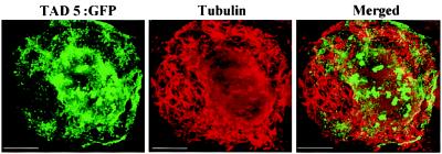
Figure 5.
TAD5:GFP does not colocalize with MTs. Protoplasts infected with TAD5:GFP were fixed and processed for immunofluorescence by using antitubulin antibody followed by a secondary antibody labeled with TRITC. The cytoplasmic filaments of tubulin (red) did not colocalize with TAD5:GFP (green). Merging the images demonstrates absence of colocalization between the signals (merged image). (Scale bar: 2.5 μm.)

