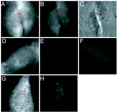
Fig. 3.
Mature miR-206 probe hybridizes in a different pattern than a probe to premiR-206, and hybridization is RNase-sensitive. Dual color in situ hybridization was performed by using a cy3-labeled LNA probe to miR-206 (A) and a fluorescein-labeled LNA probe to premiR-206 (B). (C) Phase contrast image. Red arrows point to the nucleoli. (D and E) Control using cy3-labeled LNA miR-206 probe alone. (D) Red channel. (E) Green channel. This image shows a nucleus where the nucleolar:nucleoplasmic ratio of miR-206 is ≈1:1. (F) Cy3-labeled scrambled LNA probe. A, D, and F are scaled the same, and B and E are scaled the same. (G) Fixed cells were incubated with RNase buffer alone before in situ hybridization with cy3-labeled miR-206 LNA probe. (H) Cells were treated with a mixture of three ribonucleases (see Materials and Methods) before in situ hybridization with cy3-labeled miR-206 LNA probe. G and H are scaled the same. All images are 35 μm wide.

