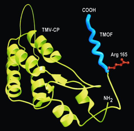
Fig. 2.
Ribbon drawing by means of MOLSCRIPT of TMV coat protein (CP) fused to A. aegypti TMOF. The TMV-CP is shown in yellow, and the N terminus is shown as NH2. TMOF exhibiting a left-handed helix is shown in blue with a trypsin cleavage site at Arg165 in red. The C terminus of the fusion protein is at the carboxyl end of TMOF (COOH).

