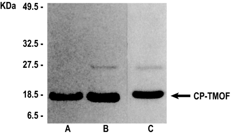
Fig. 3.
Western blot analysis and SDS/PAGE of CP-TMOF. Purified TMV-TMOF virions (22 μg, lane A; 28 μg, lane B) were run on SDS/PAGE, blotted to a membrane, and analyzed by antiserum against TMV-CP. (Lane C) Wild-type TMV-CP virions (28 μg) were run on SDS/PAGE and stained with Coomassie brilliant blue for comparison. Arrow indicates the comigration of the CP (17.5 kDa) and CP-TMOF (18.5 kDa).

