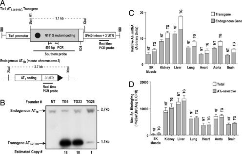
Fig. 1.
General characterization of the Tie1-AT1R TG mice. (A) Transgene construct that was injected in to mouse oocytes. The 735-bp-long EC-specific mouse Tie1 promoter, AT1R synthetic gene in which the codon for the residue Asn111 was mutated to Gly111, the 1D4 epitope tag, and the intron+ 3′ UTR of the SV40 T antigen are depicted. The C-terminal 1D4 epitope distinguishes transgene AT1R from the endogenous AT1R (15). A representation of restriction map of the mouse AT1b gene is shown. The sites for binding genotyping primers, Southern probe, and transgene transcript-specific real-time PCR probe are shown with the site of binding for the real-time PCR probe, which is specific for native AT1R mRNAs. (B) Southern blot analysis of genomic DNA digested with BamHI and XbaI from nontransgenic (NT) and transmitting TG founder mice. The probe hybridizes to 2.7- and 1.1-kb fragments, which represent the endogenous AT1b gene and the transgene, respectively. The XbaI–BamHI fragment of the AT1a gene (5.9 kb) does not appear on the gel shown. (C) Quantitative real-time PCR estimation of levels of total AT1R mRNA and endogenous AT1R mRNA (shaded fraction) in poly(A)+ RNA isolated from TG23+/− mice compared with NT. (D) Maximum specific binding (Bmax) of the 125I-[Sar1,Ile8]Ang II in membranes purified from various tissues from TG23+/− mice compared with NT mice. The shaded fraction in each bar represents the AT1R-selective binding in each tissue; the remaining binding may be caused by the AT2 receptor. Error bars represent SEM. (n >3 for each tissue). SK Muscle, skeletal muscle.

