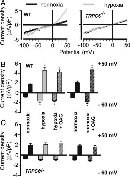
Fig. 3.
Hypoxia-induced activation of TRPC6-mediated cationic current. (A) Representative traces of whole-cell currents in normoxic and hypoxic PASMC from WT (Left) and TRPC6-deficient (TRPC6−/−, Right) mice. (B and C) Summarized data of normalized currents elicited at a potential of −60 mV and +50 mV in normoxia (black bars) and after perfusion with hypoxic bath solution (gray bars) as well as after addition of the membrane-permeable analogue of diacylglycerol (OAG), during hypoxia (gray hatched bars; n = 7 for WT and n = 11 for TRPC6−/− cells) or normoxia (black hatched bars; n = 4 each). Cells were primed with ET-1 2 min before treatment. Nifedipine was present throughout the experiments. ∗, P < 0.05 in comparison with normoxia.

