Fig. 2.
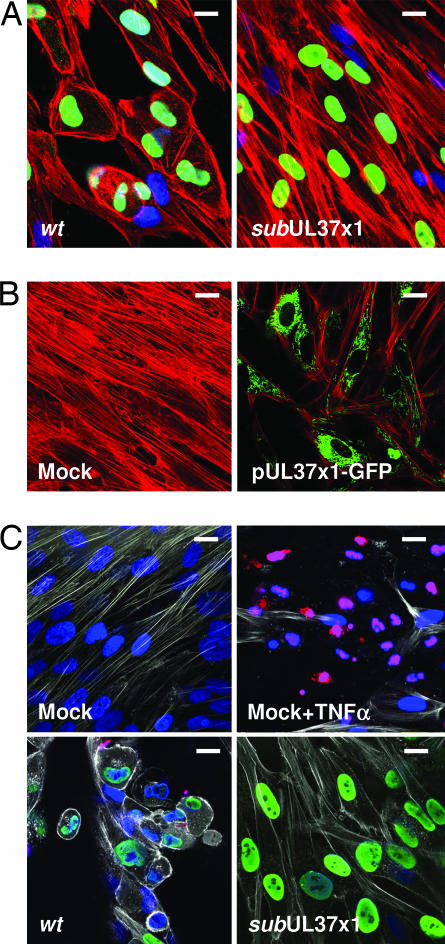
F-actin is reorganized by pUL37x1. Fibroblasts were infected at a multiplicity of ≈1 pfu per cell. (A) Immunofluorescence at 24 hpi with BADwt or BADsubUL37x1 using Alexa Fluor 546 phalloidin to detect actin (red), antibody to HCMV IE1 protein (green), and DAPI to stain DNA (blue). (B) Immunofluorescence at 24 h after mock treatment or electroporation of fibroblasts with a plasmid expressing pUL37x1-GFP using Alexa Fluor 546 phalloidin to detect actin (red) and monitoring GFP fluorescence (green). (C) TUNEL assay for apoptosis after mock infection, after treatment of mock-infected cells with TNFα, or 3 days after infection at a multiplicity of 3 pfu per cell with wt or subUL37x1. Actin was detected with Alexa Fluor 633 phalloidin (white), and IE1 protein (green) was detected by using an antibody. TUNEL-positive cells and cell fragments are red, and DNA was stained with DAPI (blue). (Scale bars: ≈10 μm.)

