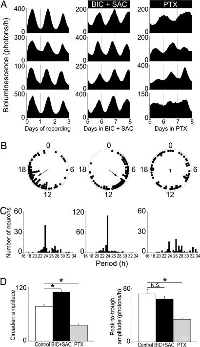
Fig. 2.
PTX, not GABA receptor antagonism, disrupts rhythms and synchrony of PER2::LUC rhythms in SCN neurons. (A) Representative PER2::LUC traces from individual neurons within mouse SCN slices under control conditions (Left), on days 5–8 of treatment with BIC+SAC (Center), and on days 5–8 of treatment with PTX (Right). (B) Representative Rayleigh plots of all rhythmic neurons within a SCN slice from each treatment group. The bioluminescence acrophase of each neuron (filled circles) and the mean phase of all neurons (arrow) are plotted for the last 24 h of each treatment. The arrow length is proportional to the magnitude of the phase clustering (r), ranging from 0 (randomly phased) to 1 (peaking at the same time). Whereas control and BIC+SAC-treated neurons maintained phase synchrony (n = 89, r = 0.63, and n = 85, r = 0.56, respectively; P < 0.001, Rayleigh test), PTX-treated neurons peaked randomly (n = 73, r = 0.16; P > 0.1). (C) Period distributions for all neurons recorded in each treatment group. PTX significantly broadened the period distribution of neurons compared with control and BIC+SAC-treated neurons (P < 0.00005, Brown–Forsythe and Levene test). (D) BIC+SAC increased, and PTX decreased the daily precision (as measured by circadian amplitude) of PER2::LUC bioluminescence rhythms in individual SCN neurons relative to controls (*, P < 0.05, ANOVA with Scheffé post hoc test). (E) Compared with controls, PTX decreased (P < 0.05), and BIC+SAC (P > 0.05) did not affect, the peak-to-trough amplitude of bioluminescence rhythms.

