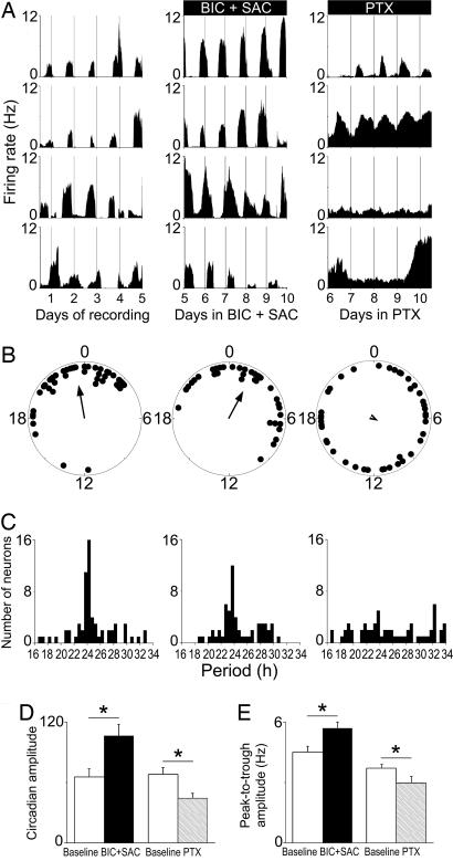
Fig. 3.
PTX, not GABA receptor antagonism, impairs firing rhythmicity and synchrony between neurons. (A) Representative firing-rate rhythms for individual SCN neurons within a control culture (Left), over the last 5 days of a 10-day BIC+SAC treatment (Center), and on the last 5 days of a 10-day PTX treatment (Right). Neurons reliably peaked at similar times during control and BIC+SAC treatment, but they were less likely to maintain rhythms, they showed lower-amplitude rhythms, and they had unstable phase relationships during PTX treatment. (B) Peak phases of all rhythmic neurons within representative SCN cultures on the last day of baseline recording (Left), on the 10th day of BIC+SAC treatment (Center), and on the 10th day of PTX treatment (Right). Rayleigh distributions of peak phases before treatment (n = 45, r = 0.66) and during BIC+SAC treatment (n = 37, r = 0.59) were statistically nonrandom (P < 0.001), but they were random during PTX treatment (n = 41, r = 0.09; P > 0.6). (C) Period distributions for all rhythmic neurons were similar before (Left) and during (Center) BIC+SAC treatment (P > 0.4), and they were significantly broadened by PTX (Right; P < 0.00001). (D) Circadian amplitudes were greater during BIC+SAC treatment than during baseline recording (*, P < 0.05) and reduced during PTX treatment relative to baseline (P < 0.05). (E) As a result of increased daily peak firing rate (P < 0.05), the peak-to-trough amplitude increased significantly during BIC+SAC treatment (P < 0.05). As a result of increased daily minimum firing rate (P < 0.00001), the peak-to-trough amplitude decreased significantly during PTX treatment (Right; P < 0.05).

