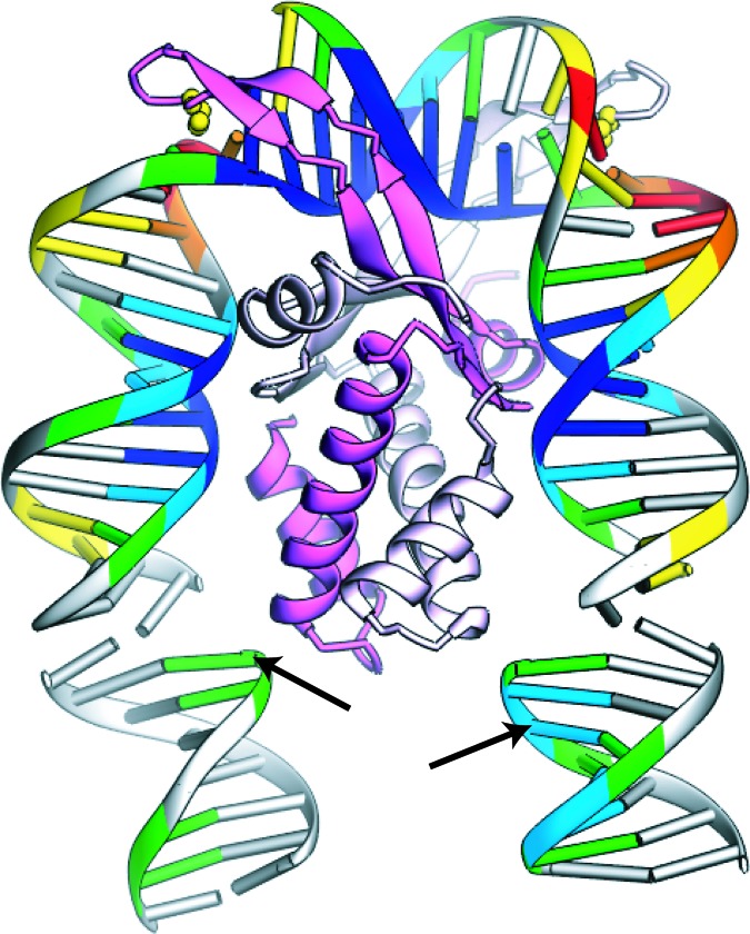
Fig. 1.
A depiction of the IHF–DNA crystal structure (17) showing the protein subunits in pink (β) and white (α) and the intercalating prolines in yellow. The DNA is color-coded according to the solvent-accessible surface revealed by the hydroxyl radical footprint in solution (5, 14): green, light blue, and dark blue indicate mild, moderate, and strong protection from cleavage, respectively, and yellow, orange, and red indicate mild, moderate, and strong enhancement of cleavage, respectively. Two segments of DNA from symmetry-related complexes within the crystal are included at the bottom to accommodate the full IHF footprint. The attachment positions for the donor and acceptor fluorophores are indicated by arrows.

