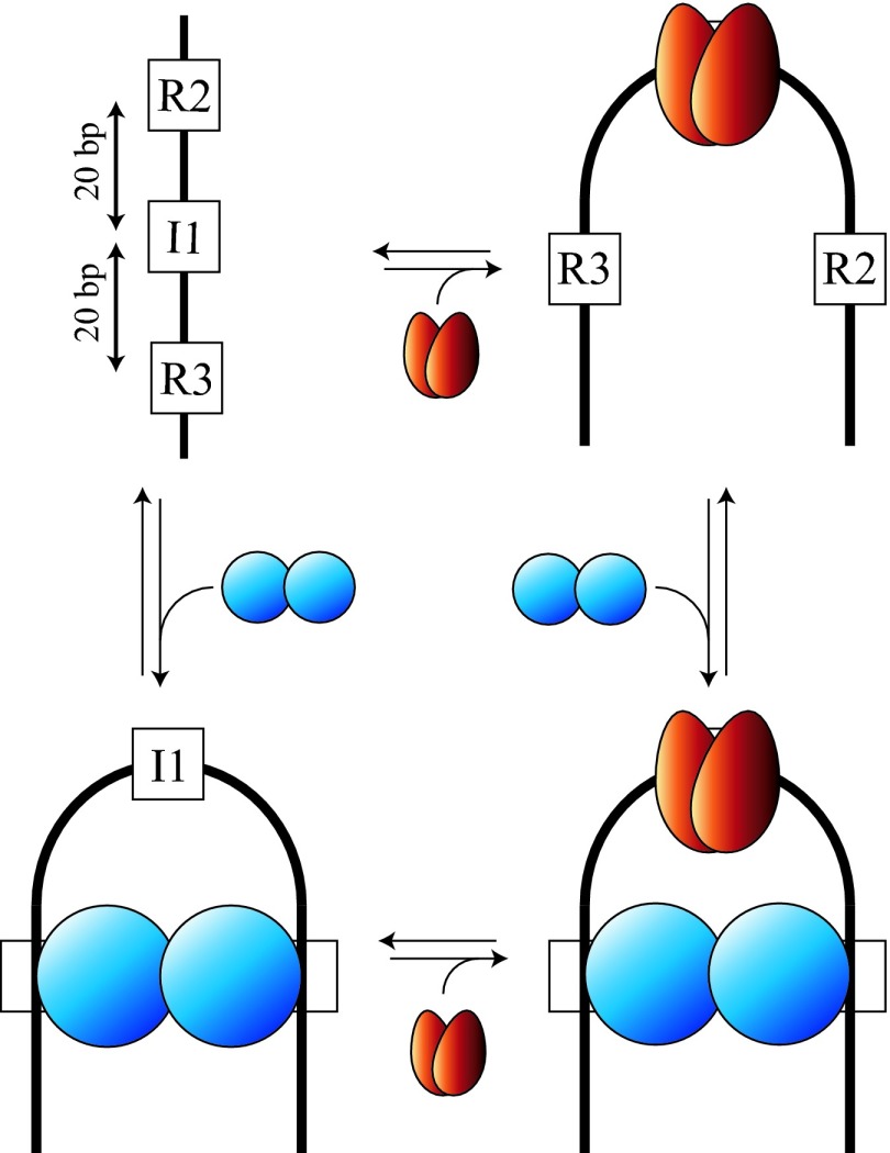
Fig. 2.
Model for the cooperative binding and bending of IHF and gpNu1 to their specific sites within cos (Upper Left; I1 and R3/R2, respectively). Bending of the duplex is induced by binding of an IHF heterodimer (red) to I1 and/or by binding of a gpNu1 homodimer (blue) to the R3 and R2 half-sites. Binding of each protein is facilitated by prebending of the duplex. (Adapted from ref. 9.)

