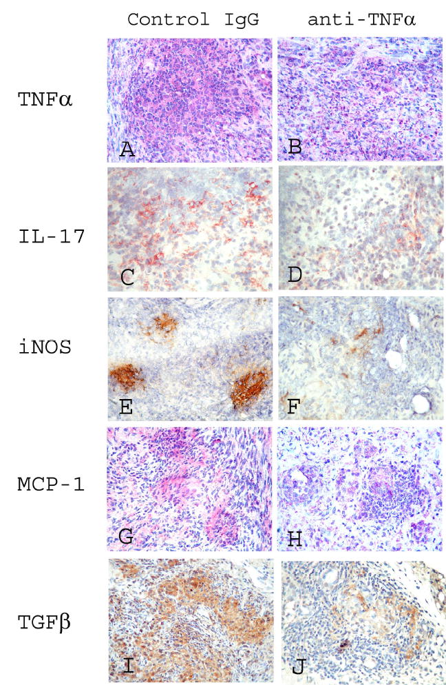
Figure 5.

Protein expression of TNFα, IL-17, iNOS, MCP-1 and TGFβ in thyroids of rat Ig-treated and anti-TNFα-treated mice. A-H: representative areas of photomicrographs demonstrating TNFα (A, B: red), IL-17 (C, D: red), iNOS (E, F: brown), MCP-1 (G, H: red), and active TGFβ (I, J: brown) at day 19 in thyroids of rat Ig-treated mice (A, C, E, G, I) and anti-TNFα-treated mice (B, D, F, H, J). G-EAT severity was 4–5+ at day 19 in thyroids of both rat Ig-treated and anti-TNFα-treated mice. Thyroids of mice given rat Ig-G had 4–5+ severity scores and fibrosis at day 40, but G-EAT lesions were resolving with little fibrosis at day 40 in thyroids of anti-TNFα-treated mice. Magnification: A–H: 400×.
