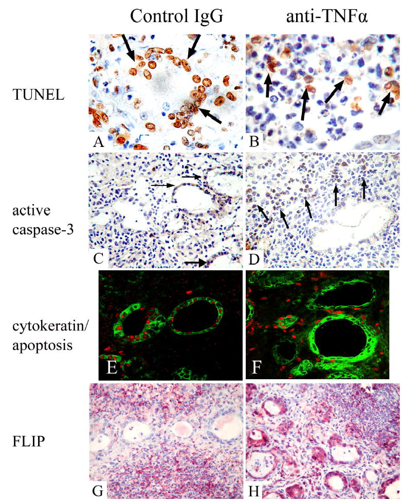
Figure 6.

Expression of apoptotic and anti-apoptotic molecules in thyroids of mice given rat IgG and anti-TNFα 19 days after cell transfer. A, B: TUNEL staining (brown) in thyroids of rat Ig-treated (A) and anti-TNFα treated mice (B). C, D: Expression of active caspase-3 (reddish brown) in thyroids of rat Ig-treated (C) and anti-TNFα-treated mice (D). E, F: Confocal analysis demonstrates apoptosis of cytokeratin+ thyrocytes in thyroids mice given rat Ig-G (E) and anti-TNFα (F). Thyrocytes were identified by cytokeratin (green, cytoplasmic staining in E and F), and apoptosis (red, nuclear staining in E, F) was detected using an in situ cell death kit. G, H: Expression of FLIP (red) in thyroids of rat Ig-treated (G) and anti-TNFα-treated mice (H). All thyroids had 4–5+ severity scores. Original magnification: A, B: 1000×; C, D, G and H: 400×; E and F, 600×.
